Tło historyczne
Na początku XX wieku Albert Einstein opracował teorię wzmocnienia światła poprzez wymuszoną emisję promieniowania (ang. Light Amplification by Stimulated Emission of Radiation, LASER). Przez 50 lat fizycy i inni naukowcy rozwijali podstawy teoretyczne, aż wreszcie w 1960 roku skonstruowano pierwszy laser.
W połowie lat 60. technologia laserowa została zastosowana przez lekarzy, którzy wierzyli, że laser może generować korzystne zmiany na poziomie komórkowym.
Dzięki badaniom klinicznym DRS i ETDRS fotokoagulacja laserowa stała się podstawową metodą leczenia retinopatii cukrzycowej od chwili jej opracowania. Wraz z powstaniem terapii anty-VEGF rola terapii laserowej zmniejszyła się, jednak od tamtej pory opracowano wiele udoskonaleń tej technologii. Od lasera termicznego do podprogowej terapii laserowej.
The very beginning
Another important time
The third important time
Na czym polega leczenie laserowe techniką SubLiminal?
Zastosowanie: wyeliminowanie jatrogennych uszkodzeń spowodowanych konwencjonalną fotokoagulacją plamki żółtej
Technika subliminal stanowi alternatywę dla konwencjonalnego lasera o fali ciągłej w leczeniu zaburzeń siatkówki i plamki żółtej. W odróżnieniu od konwencjonalnego lasera, laser podprogowy nie wywołuje uszkodzenia cieplnego siatkówki. Ma to szczególne znaczenie, gdy leczenie prowadzi się w pobliżu dołka.
Wykazano, że taka terapia laserowa jest bezpieczna i skuteczna w leczeniu patologii plamki żółtej, takich jak centralna surowicza chorioretinopatia (CSC) i cukrzycowy obrzęk plamki (DME).
Jaka długość fali?
Fotokoagulacja wymaga absorpcji światła przez określone pigmenty w oku.
Trzy główne pigmenty w oku to melanina, hemoglobina i ksantofil, które pochłaniają różne długości fal świetlnych i występują w różnych proporcjach w siatkówce.
Teoretycznie żółte światło laserowe o długości 577 nm zapewnia pik absorpcji oksyhemoglobiny, doskonałą widoczność zmiany, niski stopień rozpraszania w gałce ocznej, niski poziom bólu i pomijalną absorpcję przez ksantofil1,2.
Uznaje się, że żółte światło laserowe o długości 577 nm jest najbezpieczniejsze i najbardziej uniwersalne3.
Thanks to its absorption characteristics, it causes lesser scatter and requires the use of a lower energy level compared to green (532nm) and other yellow (561 to 568nm) wavelengths3.
It is minimally absorbed by macular xanthophylls45, potentially allowing for treatments close to the fovea6.
It is highly absorbed by the oxyhemoglobin (absorption peak) and thus optimal for the treatment of vascular lesions and subretinal vascular proliferations7.
Jak przebiega leczenie laserowe techniką SubLiminal?
Leczenie laserowe techniką SubLiminal jest nowoczesną techniką lasera podprogowego, która obejmuje spersonalizowaną siatkę wzorców i możliwość dostarczania energii lasera w krótkich, mikrosekundowych impulsach.
Umożliwia to schłodzenie nabłonka barwnikowego siatkówki (RPE) pomiędzy impulsami, co zapobiega skumulowaniu krytycznego poziomu ciepła w tkance, a w rezultacie bliznowaceniu RPE i siatkówki, które stanowi znaną, lecz możliwą do uniknięcia odpowiedź na to leczenie oraz ogranicza możliwość ponownego leczenia w przyszłości.
Clinical data
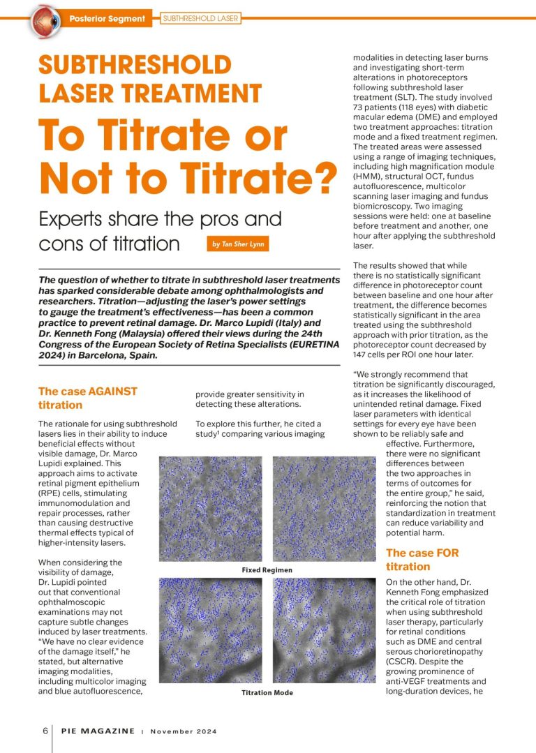
Subthreshold laser treatment: To Titrate or Not to Titrate?
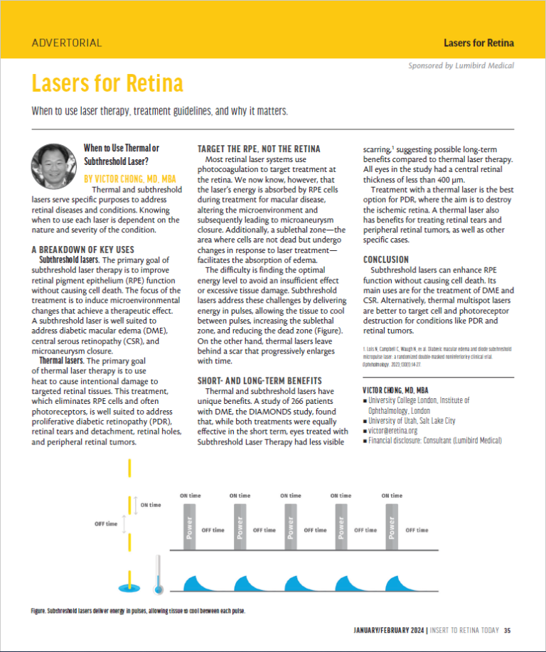
Lasers for Retina
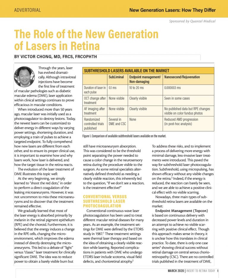
The Role of the New Generation of Lasers in Retina
Vascular endothelial growth factor inhibition and proliferative diabetic retinopathy, a changing treatment paradigm?
How new generation lasers are different from each other
Comparison of 577-nm Multispot and Standard Single-Spot Photocoagulation for Diabetic Retinopathy
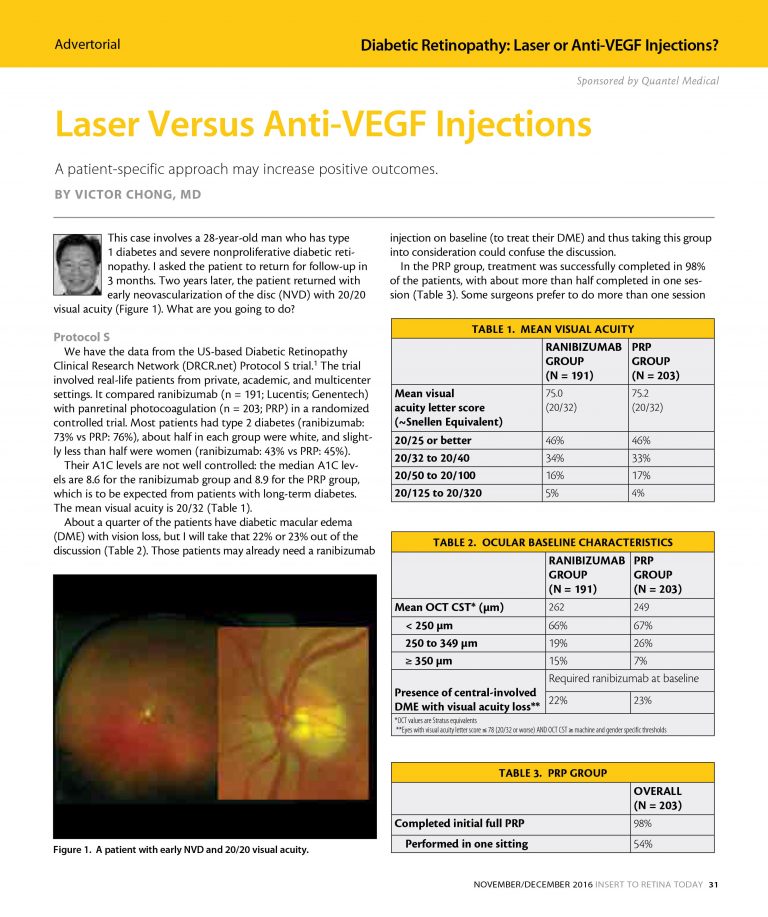
Laser Versus Anti-VEGF Injections
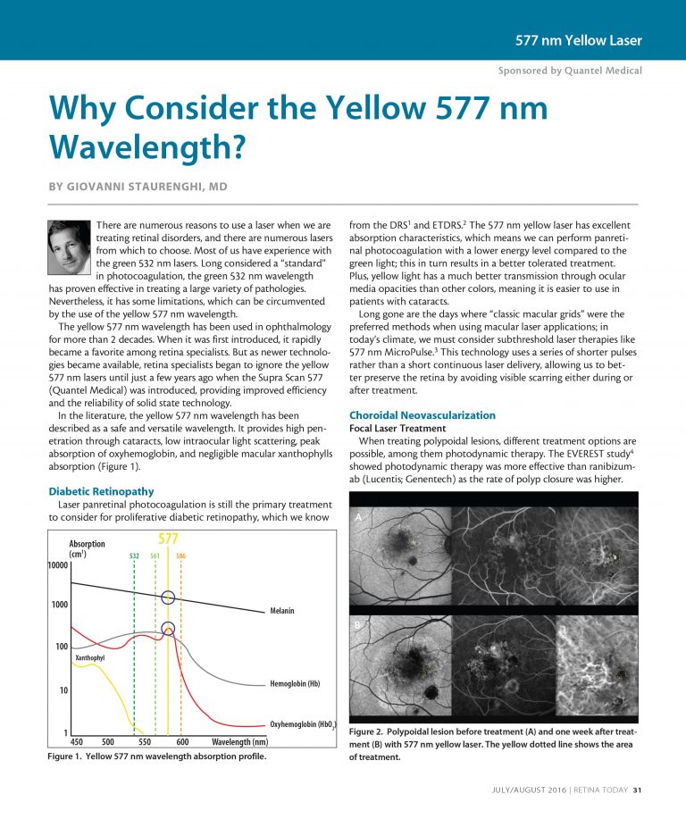
Why Consider the Yellow 577 nm Wavelength?
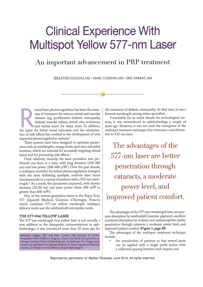
Clinical experience with Multispot yellow 577nm laser
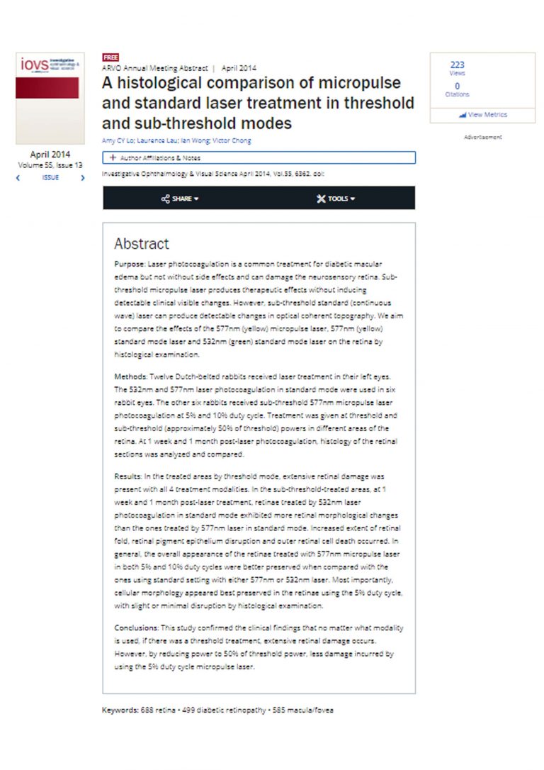
A histological comparison of micropulse and standard laser treatment in threshold and sub-threshold modes
Do We Still Need Subthreshold Laser for Macular Diseases?
How to optimize retina laser treatments in 2020?
Subliminal laser treatment for macular diseases
How are retina lasers different from each other ?
Controversies on Modern Laser Treatments for Retina and Vitreous
SubLiminal Laser Therapy: Concept and reasons
Expanding clinical application of SubLiminal laser
Why consider the yellow 577nm wavelength?
Bibliografia
Graefes Arch Clin Exp Ophthalmol. 1989;227:277-280.
Ophthalmology.1986;93:952-958
Purpose: This pilot study aimed to evaluate the efficacy and safety of subthreshold micropulse yellow (577-nm) laser photocoagulation (SMYLP) in the treatment of diabetic macular edema (DME).
Methods: We reviewed 14 eyes of 12 patients with DME who underwent SMYLP with a 15% duty cycle at an energy level immediately below that of the test burn. The laser exposure time was 20 ms and the spot diameter was 100 µm. Laser pulses were administered in a confluent, repetitive manner with a 3 × 3 pattern mode.
Results: The mean follow-up time was 7.9 ± 1.6 months. The baseline-corrected visual acuity was 0.51 ± 0.42 logarithm of the minimum angle of resolution (logMAR), which was improved to 0.40 ± 0.35 logMAR (p = 0.025) at the final follow-up. The central macular thickness at baseline was 385.0 ± 111.0 µm; this value changed to 327.0 ± 87.7 µm (p = 0.055) at the final follow-up.
Conclusions: SMYLP showed short-term efficacy in the treatment of DME and did not result in retinal damage. However, prospective, comparative studies are needed to better evaluate the efficacy and safety of this treatment.
Keywords: Diabetic retinopathy; Laser therapy; Macular edema.
J Ophthalmol. 2015.
